Welcome to this comprehensive guide designed to demystify the term “graphy” within medical terminology. Whether you’re a medical student, a healthcare professional, or simply someone who is curious about medical jargon, this guide will break down the concept of “graphy” and its applications in healthcare. By following this step-by-step guidance, you’ll gain a robust understanding and practical knowledge that you can apply directly in real-world scenarios.
Understanding Graphy in Medical Terms: Your Complete Guide
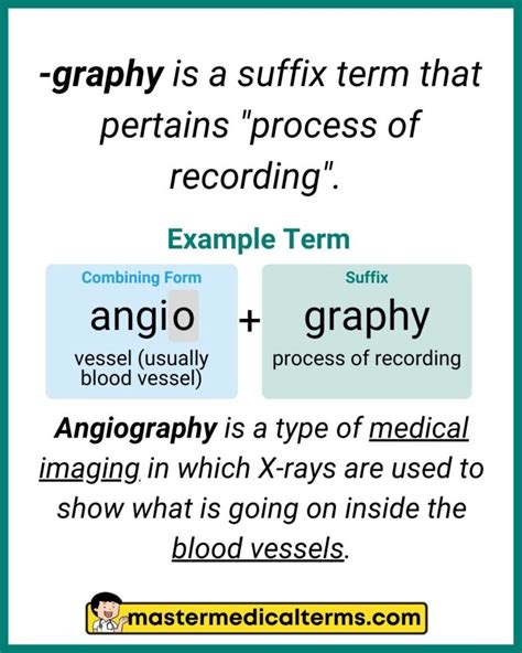
When we delve into medical terminology, we often encounter terms that, at first glance, seem daunting. One such term is “graphy.” Simply put, “graphy” is a suffix that refers to the process of recording or imaging. It’s a cornerstone of many medical terms you’ll encounter in your healthcare journey.
Imagine this: you’re diagnosing a patient, and various imaging techniques come into play. Understanding that “graphy” often denotes the recording of images, such as X-rays, MRIs, or CT scans, will instantly make a lot more sense. This guide aims to unravel this concept and illustrate its practical applications.
Quick Reference
Quick Reference
- Immediate action item: Start by familiarizing yourself with common medical imaging techniques.
- Essential tip: Understand the prefix associated with “graphy” to quickly decipher medical terms.
- Common mistake to avoid: Confusing “graphy” with “gram” (which refers to a recorded image, not the process of recording).
To make this knowledge stick, let’s dive into more detailed how-to sections that cover the foundational to advanced aspects of graphy in healthcare.
How to Identify and Understand Medical Grafhy Terms
Medical terminology often uses “graphy” to denote various imaging processes. To better understand this, let’s break it down step-by-step.
First, consider the following:
- X-ray radiography: The process of producing images of the internal structures of the body using X-rays.
- Ultrasonography: The use of ultrasound waves to produce images of structures within the body.
- Magnetic resonance imaging (MRI): A technique that uses magnetic fields and radio waves to produce detailed images of the organs and tissues within the body.
Understanding these terms begins with recognizing “graphy” as your key indicator. This suffix tells us that the process involves creating visual records of the internal anatomy. Now let’s look at a more in-depth breakdown:
Step-by-Step Identification:
- Identify the Suffix: Look for “-graphy” at the end of medical terms to find imaging processes.
- Analyze the Prefix: Examine the prefix to determine the specific body part or type of imaging involved.
- Combine and Decipher: Combine the prefix and suffix to understand the complete term and its application.
By following these steps, you can quickly decipher complex medical terms related to graphy.
Mastering the Applications of Graphy in Diagnostics
Now that we’ve covered the basics, let’s explore the practical applications of graphy in medical diagnostics.
Application in Radiology: Radiologists use various graphy techniques to diagnose and treat conditions. For instance, computed tomography (CT) scans utilize X-rays to produce cross-sectional images of the body. These scans are crucial for identifying internal injuries, tumors, or other abnormalities.
Application in Cardiology: In cardiology, techniques like echocardiography use ultrasound waves to produce images of the heart. This helps in diagnosing heart conditions, such as valve disorders or heart enlargement.
Application in Neurology: Neurologists employ magnetic resonance imaging (MRI) to visualize the brain and spinal cord. MRI provides detailed images of brain structures, helping to diagnose neurological conditions like multiple sclerosis, brain tumors, or stroke.
Here’s a simple framework to apply graphy techniques in diagnostics:
- Select Appropriate Technique: Choose the imaging method that best suits the diagnostic needs.
- Patient Preparation: Ensure the patient is prepared for the procedure. This may include fasting, removing metal objects, or taking specific medications.
- Perform the Procedure: Follow the protocol for the chosen graphy technique, ensuring proper positioning and timing.
- Interpret Results: Analyze the images and compare them with normal anatomical standards to identify any abnormalities.
Practical FAQ Section
What is the difference between “graphy” and “gram” in medical terms?
While both terms involve imaging, the key difference lies in their use. “Graphy” refers to the process of creating an image, whereas “gram” refers to the actual recorded image itself. For example, in ultrasonography, the process is referred to as ultrasonography, while the recorded image is an ultrasound gram.
How do I ensure accurate results in medical imaging?
To ensure accurate results in medical imaging, follow these steps:
- Patient Communication: Clearly communicate with the patient to understand any medical history or conditions that might affect the imaging process.
- Technique Proficiency: Ensure that the operator is well-trained and proficient in the specific imaging technique.
- Quality Control: Regularly calibrate the imaging equipment to maintain high-quality standards.
- Image Analysis: Carefully analyze the images for any abnormalities, cross-referencing with the patient’s medical history.
Can imaging techniques be harmful?
While most imaging techniques are safe, some, like CT scans and X-rays, use ionizing radiation, which carries a small risk of increasing cancer risk. However, the benefits generally outweigh the risks, especially when used for diagnostic purposes. It’s important to use the lowest possible dose needed for accurate diagnosis and to follow guidelines for safe exposure. Non-ionizing techniques like ultrasound and MRI do not carry the same risks.
By grasping the fundamental concept of “graphy” and understanding its practical applications, you’ll be well-equipped to navigate the world of medical imaging. This guide has provided you with the knowledge and tools to identify and apply various graphy techniques in real-world medical scenarios. Keep practicing and exploring to continually enhance your expertise.