Welcome to Heart Model Labeled: Anatomy Unveiled
Understanding the anatomy of the heart is crucial for anyone interested in cardiovascular health, medicine, or simply for general knowledge. Navigating through the complexities of this vital organ can be daunting, but this guide aims to simplify the process, offering step-by-step guidance with actionable advice. From basic concepts to advanced understanding, this guide will take you through the heart’s anatomy in a practical, user-friendly manner.
Whether you’re a student, a healthcare professional, or simply curious, this guide covers everything you need to know about the heart’s structure and function. We’ll address common pain points in learning about heart anatomy and provide practical examples and solutions to make the information accessible and useful. Ready to dive in? Let's get started!
Problem-Solution Opening Addressing User Needs
Many people find the heart’s anatomy intimidating due to its complex structure and the jargon often used in medical literature. This guide is designed to break down these complexities into understandable and actionable pieces of information. Whether you’re struggling to grasp the basics of heart chambers, valves, and vessels, or seeking to understand more advanced concepts like electrical conduction pathways, this guide provides clear, practical advice to meet your needs. We’ll focus on real-world applications and examples that you can implement immediately, making the learning process efficient and engaging.
Quick Reference
Quick Reference
- Immediate action item: Start with identifying the four chambers of the heart and the main arteries and veins connected to them for a foundational understanding.
- Essential tip: Use mnemonic devices or visual aids like labeled diagrams to better remember the intricate pathways of blood flow through the heart.
- Common mistake to avoid: Confusing the right and left sides of the heart; ensure to understand the unique features and functions of each side.
The Basics of Heart Anatomy
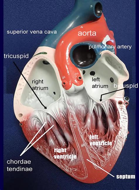
Let’s begin with the basics: understanding the four chambers of the heart is your first step towards mastering heart anatomy. The heart is divided into four chambers: the left atrium, right atrium, left ventricle, and right ventricle. Each chamber plays a critical role in the circulatory system.
Left Atrium: This chamber receives oxygen-rich blood from the lungs via the pulmonary veins. Its primary function is to collect this oxygenated blood and pump it into the left ventricle.
Right Atrium: The right atrium collects oxygen-poor blood from the body through the superior and inferior vena cava. Its main role is to prepare the blood for oxygenation in the lungs.
Left Ventricle: The powerhouse of the heart, the left ventricle pumps oxygenated blood into the aorta, distributing it throughout the body.
Right Ventricle: This chamber sends oxygen-poor blood to the lungs via the pulmonary artery, where it gets oxygenated.
To better understand the movement of blood through the heart, consider the following steps:
- Oxygen-poor blood from the body enters the right atrium.
- It moves to the right ventricle and is pumped to the lungs.
- Oxygen-rich blood returns to the left atrium from the lungs.
- It passes into the left ventricle, which then pumps it out to the body.
This cycle is powered by the heart’s electrical conduction system, which we’ll cover later in more detail.
Navigating the Heart’s Electrical System
Understanding the heart’s electrical system is essential for grasping how it coordinates contractions and keeps your heart beating efficiently. The system comprises several key components:
- Sinoatrial (SA) Node: Known as the heart’s natural pacemaker, the SA node initiates the electrical impulses that trigger the heartbeat.
- Atrioventricular (AV) Node: This node delays the signal before it moves from the atria to the ventricles, allowing the atria to contract fully before the ventricles.
- Bundle of His: This pathway transmits the electrical signal from the AV node into the ventricles.
- Purkinje Fibers: These fibers spread the signal throughout the ventricles, triggering their contraction.
To visualize and remember this pathway, draw a diagram and label each component. Follow this with a simple mnemonic: “SA Node kicks off the race, AV Node gives it a pause, Bundle of His sends the lead, Purkinje fibers finish the chase.”
Deep Dive into the Heart’s Valves
The heart’s valves are crucial for ensuring one-way blood flow. There are four main valves in the heart:
- Tricuspid Valve: Located between the right atrium and right ventricle, it prevents backflow of blood as the right ventricle contracts.
- Pulmonary Valve: Guards the exit from the right ventricle to the pulmonary artery, stopping blood from flowing back when the right ventricle relaxes.
- Mitral Valve (or Bicuspid Valve): Between the left atrium and left ventricle, it ensures blood doesn’t flow back into the left atrium.
- Aortic Valve: Located between the left ventricle and the aorta, it prevents blood from flowing back into the left ventricle after it has been pumped into the aorta.
Understanding the function of these valves is critical for diagnosing and treating heart conditions. When valves don’t function correctly, it can lead to issues like regurgitation (backflow of blood) or stenosis (narrowing of the valve opening). To prevent these problems, it’s essential to maintain heart health through proper diet, exercise, and regular check-ups.
Practical Tips for Mastering Heart Anatomy
Here are some practical tips to help you master heart anatomy efficiently:
- Use labeled heart models and diagrams to get a hands-on and visual understanding.
- Regularly test your knowledge by creating flashcards or quizzes with terms and functions.
- Watch educational videos that visually demonstrate the heart’s structure and function.
Practical FAQ
What are the most common diseases affecting the heart?
Several common diseases can affect the heart, including coronary artery disease (CAD), heart failure, arrhythmias, and congenital heart defects. CAD occurs when the arteries that supply blood to the heart become narrowed or blocked. Heart failure happens when the heart cannot pump enough blood to meet the body’s needs. Arrhythmias involve irregular heartbeats, which can be too fast, too slow, or uncoordinated. Congenital heart defects are present from birth and can affect the structure of the heart. Understanding these conditions can help in recognizing symptoms and seeking timely medical attention.
Advanced Insights into Heart Anatomy
For those looking to delve deeper into heart anatomy, consider the following advanced insights:
- Microanatomy: Examine the cellular structure of the heart, including the types of tissues that make up the chambers and valves.
- Functional Histology: Study the microscopic components like cardiac muscle fibers, connective tissue, and blood vessels that constitute the heart.
- Surgical Anatomy: Learn about the anatomical landmarks relevant to cardiac surgery, such as the positions of major vessels and the heart’s proximity to other thoracic organs.
Understanding these aspects requires more detailed study and often the use of advanced imaging techniques like CT scans or MRIs. However, this knowledge can be invaluable for professionals in cardiac surgery and interventional cardiology.
Conclusion
Demystifying the heart’s anatomy can seem overwhelming at first, but with the right approach and resources, it becomes an achievable and fascinating subject to master. This guide aims to provide a comprehensive, step-by-step understanding, starting from the basics and moving into more advanced topics. Remember, practical application and real-world examples are key to truly grasping complex concepts. Keep practicing, stay curious, and don’t hesitate to seek out additional resources as needed. Happy learning!